Vascular Lesions

Book a Consultation

Fill in the form below to get started.

What are Vascular Lesions?

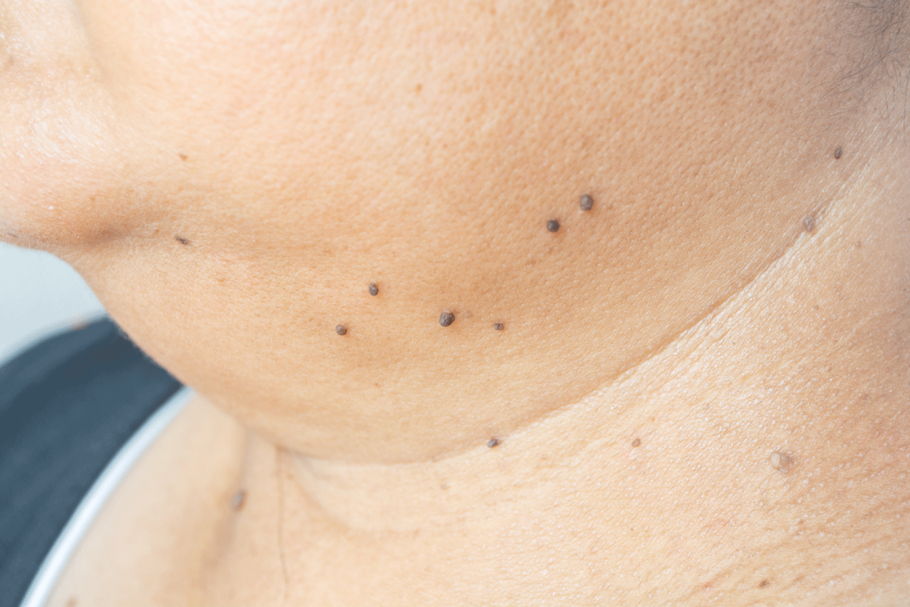
Vascular lesions are abnormalities in blood vessels, including veins, arteries, and capillaries, that can be either congenital or acquired. Subtypes include haemangiomas, which are benign tumors composed of blood vessels often appearing as red or purple marks on the skin, and angiomas, a broader category encompassing benign growths such as cherry angiomas (small, red, dome-shaped lesions) and spider angiomas (central red spots with radiating blood vessels).

Types of Vascular Lesions

There are different types of vascular lesions including: 

Infantile Haemangiomas:

  • Symptoms: Red or purple raised lesions on the skin, typically appearing within the first few weeks of life. They may grow rapidly before gradually shrinking.
  • Characteristics: Usually benign and self-resolving, but can occasionally cause complications if located near vital structures.


Cavernous Haemangiomas:

  • Symptoms: Blue or purple spongy masses under the skin, often deeper than infantile haemangiomas.
  • Characteristics: Can cause pain or functional impairment depending on their size and location.

Cherry Angiomas:

  • Symptoms: Small, bright red, dome-shaped lesions on the skin, commonly found on the trunk and limbs.
  • Characteristics: Typically asymptomatic and benign; they may bleed if scratched.


Spider Angiomas:

  • Symptoms: Central red spot with radiating blood vessels, resembling a spider’s web, often found on the face, neck, and upper body.
  • Characteristics: Generally benign but may be associated with liver disease or hormonal changes.
  • Symptoms: Flat, pink, red, or purple birthmarks that typically appear on the face, arms, or legs.
  • Characteristics: Permanent and may darken over time; can be associated with syndromes such as Sturge-Weber.
  • Symptoms: Soft, compressible blue masses, often becoming more prominent with age.
  • Characteristics: Can cause pain, swelling, and functional impairment depending on size and location.
  • Symptoms: Pulsatile masses, skin warmth, and overlying skin color changes; may cause pain or bleeding.
  • Characteristics: Abnormal connections between arteries and veins, potentially leading to serious complications like bleeding or heart issues.

Treatment Options

There is a spectrum of size, shape and depth of vascular lesions of which each subtype may require a different treatment strategy. At Cathedral Dermatology we can treat small and superficial haemangiomas on the skin with surgery or cautery, however, if a lesion requires further complex treatment or laser, our team will recommend the relevant specialists for you to see for treatments that we do not provide at the clinic.

Infantile Haemangiomas:

  • Medications:
    • Propranolol: A beta-blocker that reduces blood flow to the haemangioma, leading to decreased size and color.
    • Corticosteroids: Oral or topical corticosteroids can help reduce inflammation and size.
  • Laser Therapy: Pulsed dye lasers can be used to target the blood vessels within the haemangioma, reducing redness and improving cosmetic appearance. Multiple sessions may be needed.
  • Surgery: Reserved for cases where haemangiomas cause significant functional impairment (e.g., obstructing vision) or cosmetic issues that do not improve with other treatments. Surgical options include excision or reconstructive techniques.


Cavernous Haemangiomas:

  • Medications:
    • Corticosteroids: Can reduce the size and symptoms of cavernous haemangiomas by decreasing inflammation and slowing blood flow.
    • Propranolol: Sometimes used if there is a significant impact on function or appearance.
  • Surgery: Recommended for larger or symptomatic lesions that cause pain, bleeding, or functional impairment. Surgical removal involves excising the lesion, which may require specialized techniques depending on its depth and location.
  • Sclerotherapy: Involves injecting a sclerosing agent (such as ethanol or polidocanol) directly into the haemangioma to induce scarring and shrink the lesion. This approach can be used alone or in combination with other treatments.

Cherry Angiomas:

  • Observation: Often benign and asymptomatic, so no treatment is required unless for cosmetic reasons.
  • Laser Therapy:
    • Pulsed Dye Laser: Targets the blood vessels in the angioma, causing them to collapse and fade. Multiple sessions may be required.
    • Nd Laser: Sometimes used for deeper lesions.
  • Cryotherapy: Involves freezing the angioma with liquid nitrogen. This method destroys the abnormal blood vessels, causing them to fall off over time.
  • Electrocautery: Uses an electric current to burn the angioma off. This procedure is quick and effective, but may leave a small scar.


Spider Angiomas:

  • Laser Therapy:
    • Pulsed Dye Laser: Targets the blood vessels, reducing the appearance of the spider angioma.
    • Nd Laser: Effective for treating more extensive or deeper spider angiomas.
  • Electrocautery: Similar to cherry angiomas, this technique uses heat to remove the spider angioma.
  • Laser Therapy:
    • Pulsed Dye Laser: The primary treatment for port-wine stains, this laser selectively targets the blood vessels within the stain, reducing redness and improving skin texture. Multiple sessions are usually required, and the treatment may cause temporary redness or swelling.
  • Camouflage Makeup: Specially formulated makeup can effectively conceal port-wine stains. Products designed for this purpose provide high coverage and are resistant to water and sweat.
  • Surgery: Rarely used but may be considered for severe cases or associated complications. Techniques may include skin grafts or reconstructive surgery.
  • Sclerotherapy:
    • Procedure: A sclerosing agent (e.g., ethanol, polidocanol) is injected into the venous malformation to irritate the lining of the veins, causing them to shrink and eventually be absorbed by the body. Multiple sessions may be required, depending on the size and complexity of the malformation.
  • Laser Therapy: Used for superficial venous malformations. Lasers target the blood vessels, causing them to collapse and improve the appearance of the skin.
  • Surgery: Involves excising large or problematic venous malformations. Surgical planning may include preoperative imaging and coordination with other treatments.
  • Compression Garments: Used to manage symptoms like swelling and discomfort. These garments apply consistent pressure to the affected area, helping to reduce swelling and improve circulation.
  • Embolization:
    • Procedure: A catheter is inserted into the blood vessels and guided to the AVM, where embolic agents (such as glue or coils) are injected to block the abnormal blood flow. This reduces the size and blood flow of the AVM, decreasing the risk of bleeding.
  • Surgery:
    • Procedure: Surgical removal of the AVM involves excising the abnormal vessels and repairing the surrounding tissues. This option is considered when the AVM is accessible and poses a significant risk if left untreated.
  • Stereotactic Radiosurgery:
    • Procedure: Focused radiation is used to target the AVM, causing the abnormal blood vessels to gradually close off. This non-invasive method requires careful planning and may take several years to achieve full results.

Book a Consultation

Fill in the form below to get started.